Recent Publications

A unifying mechanism for presynaptic homeostatic plasticity at mammalian peripheral and central synapses
Presynaptic homeostatic plasticity (PHP) is a potent form of adaptive plasticity that has been documented at synapses as diverse as the glutamatergic Drosophila neuromuscular junction (NMJ), cholinergic mammalian NMJ (including human), and glutamatergic synapses in the mammalian brain. We define secreted class III semaphorin as a unifying, trans-synaptic signal necessary for PHP at highly divergent synapses. Sema3a drives the rapid induction of PHP at the cholinergic mouse NMJ and synapses in the adult hippocampus (CA1), including cross-modal potentiation of inhibitory transmission. Read more here...
Peter H. Chipman, Unghwi Lee, Brian O. Orr, Richard D. Fetter, Graeme W. Davis, A unifying mechanism for presynaptic homeostatic plasticity at mammalian peripheral and central synapses, Neuron,2025,,ISSN 0896-6273, https://doi.org/10.1016/j.neuron.2025.05.030.
Cadherin-13 Maintains Retinotectal Synapses via Transneuronal Interactions
Abstract: Maintaining precise synaptic contacts between neuronal partners is critical to ensure the proper functioning of the mammalian central nervous system (CNS). Diverse cell recognition molecules, such as classic cadherins (Cdhs), are part of the molecular machinery mediating synaptic choices during development and synaptic maintenance. Yet, the principles governing neuron–neuron wiring across diverse CNS neuron types remain largely unknown...Continue Reading Here
Angela C. Matcham, Kenichi Toma, Nicole Y. Tsai, Christina J. Sze, Pin-Yeh Lin, Ilaria F. Stewart, Xin Duan. Journal of Neuroscience 31 January 2024, 44 (5) e1310232023; DOI: 10.1523/JNEUROSCI.1310-23.2023
Hindbrain modules differentially transform activity of single collicular neurons to coordinate movements
Abstract: Seemingly simple behaviors such as swatting a mosquito or glancing at a signpost involve the precise coordination of multiple body parts. Neural control of coordinated movements is widely thought to entail transforming a desired overall displacement into displacements for each body part. Here we reveal a different logic implemented in the mouse gaze system... Continue Reading Here
Zahler SH, Taylor DE, Wright BS, Wong JY, Shvareva VA, Park YA, Feinberg EH. Hindbrain modules differentially transform activity of single collicular neurons to coordinate movements. Cell. 2023 Jul 6;186(14):3062-3078.e20. doi: 10.1016/j.cell.2023.05.031. Epub 2023 Jun 20. PMID: 37343561; PMCID: PMC10424787.
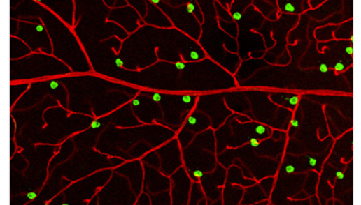
Neurons in the retina (green) making directing blood vessels (red) to form a complex, three-dimensional lattice as they grow.
Molecular and spatial analysis of ganglion cells on retinal flatmounts identifies perivascular neurons resilient to glaucoma
Abstract: Recent transcriptomic studies have categorized mouse retinal ganglion cells (RGCs) into 45 types; however, little is known about their spatial distributions on the two-dimensional retinal surface and how their local microenvironments impact their functions. Here, we optimized a workflow combining imaging-based spatial transcriptomics (multiplexed-error robust fluorescent in situ hybridization [MERFISH]) and immunostaining on retinal flatmounts. We computationally registered the somata distributions of all RGCs and found that 34/45 molecularly defined types exhibited non-uniform distributions. We analyzed local neighborhoods for each cell and identified seven RGC types enriched in the perivascular niche, including direction-selective RGC (DSGC) and intrinsically photosensitive RGC (ipRGC) types. Continue Reading...
Nimkar K, Tsai NY, Zhao M, Yi Y, Lum MR, Garrett TR, Wang Y, Toma K, Caval-Holme F, Reddy N, Ehrlich AT, Kriegstein AR, Do MTH, Hu Y, Sivyer B, Shekhar K, Duan X. Molecular and spatial analysis of ganglion cells on retinal flatmounts identifies perivascular neurons resilient to glaucoma. Neuron. 2025 Aug 19:S0896-6273(25)00552-5. doi: 10.1016/j.neuron.2025.07.025. Epub ahead of print. PMID: 40840447.
Perivascular neurons instruct 3D vascular lattice formation via neurovascular contact.
Abstract: The vasculature of the central nervous system is a 3D lattice composed of laminar vascular beds interconnected by penetrating vessels. The mechanisms controlling 3D lattice network formation remain largely unknown. Combining viral labeling, genetic marking, and single-cell profiling in the mouse retina, we discovered a perivascular neuronal subset, annotated as Fam19a4/Nts-positive retinal ganglion cells (Fam19a4/Nts-RGCs), directly contacting the vasculature with perisomatic endfeet. Continue reading here...
Perivascular neurons instruct 3D vascular lattice formation via neurovascular contact. Cell. 2024 23 May 2024, Pages 2767-2784. Toma K, Zhao M, Zhang S, Wang F, Graham HK, Zou J, Modgil S, Shang WH, Tsai NY, Cai Z, Liu L, Hong G, Kriegstein AR, Hu Y, Körbelin J, Zhang R, Liao YJ, Kim TN, Ye X, Duan X. PMID: 38733989 https://doi.org/10.1016/j.cell.2024.04.010
